 فارسی
فارسی 
Abstract
The 450 nm wavelength blue diode laser is one of the newest technologies used in soft tissue oral surgeries, which provides significant results even with minimal power. This method allows for precise surgery, without pain or bleeding, without the need for stitches, rapid recovery, and greater patient satisfaction. This article introduces the clinical application of this laser in the removal of a lower lip fibroma and examines its benefits, results, and therapeutic values.
Keywords: blue laser, diode, oral surgeries, patient satisfaction, oral soft tissue
Introduction
Since the introduction of lasers in dentistry in the 1960s, this technology has brought about major changes in the field of oral and dental treatments. Laser surgery not only reduces operating time and bleeding control, but also provides better visibility for the surgeon and a more comfortable experience for the patient due to its high precision.
Various lasers are used in oral surgeries, but blue light (450 nm) has attracted much attention due to its unique properties. In addition to its antibacterial properties and helping to repair tissues, this light has also been used in cases such as tooth whitening and polymerization of dental resins. However, its application in oral soft tissue surgery is a new and developing topic.
Materials and Methods
A 38-year-old female patient presented with a lesion on the edge of the lower lip. The lesion, which had developed over a year, had grown more in recent months. The clinical diagnosis was fibroma (benign lesion). To avoid bleeding and scarring caused by traditional surgery, lesion removal with a 450 nm diode laser was chosen.
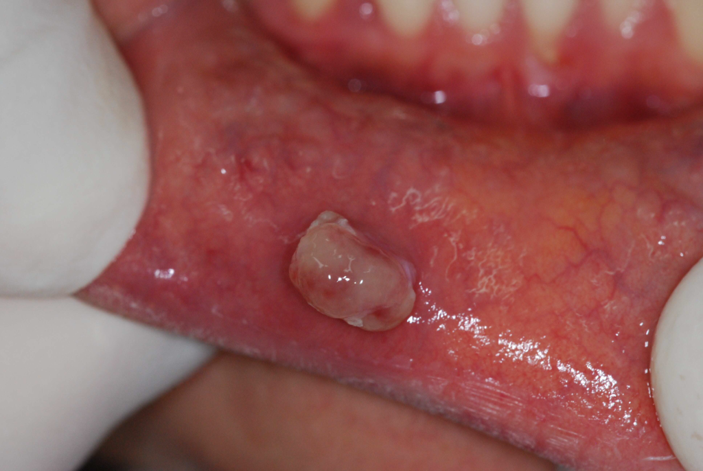
Figure 1. Lower lip fibroma, possibly caused by lip biting
Features of this procedure:
No need for anesthetic injection (local anesthesia only)
No sutures
Operation time less than two minutes
Complete control of bleeding and tissue temperature
Follow-up of the patient at one and three months after the procedure showed that no complications occurred and the lesion did not return.
Results
The patient did not feel any pain during the procedure.
After the lesion was removed, there was no bleeding and no sutures were required.
The surgical site healed completely within a week.
The patient did not take any medications such as antibiotics or painkillers during the recovery period.
No infection, swelling, or discomfort was reported.
Histopathological examination of the specimen showed fibrous connective tissue with a neat cut and minimal carbonization.
Key advantage: This laser allows for precise removal of the lesion with minimal tissue damage, and the quality of the specimen for pathological examination is fully preserved.
Resources:
- Goldman L, Gray JA, Goldman J, Goldman B, Meyer R. Effect of laser beam impacts on teeth. J Am Dent Assoc 1965; 70: 601-606 [PMID: 14245324 DOI: 10.14219/jada.archive.1965.0260]
- Kinersly T, Jarabak JP, Phatak NM, Dement J. Laser effects on tissue and materials related to dentistry. J Am Dent Assoc 1965; 70: 593-600 [PMID: 14245323 DOI: 10.14219/jada.archive.1965.0268]
- Morrant GA. Lasers: an appraisal of their possible use in dentistry. Dent Pract Dent Rec 1965; 16: 5-9 [PMID: 5212929]
- Stern RH, Sognnaes RF. Laser effect on dental hard tissues. a preliminary report. J South Calif Dent Assoc 1965; 33: 17-19 [PMID: 14241171]
- Taylor R, Shklar G, Roeber F. The effects of laser radiation on teeth, dental pulp, and oral mucosa of experimental animals. Oral Surg Oral Med Oral Pathol 1965; 19: 786-795 [PMID: 14293970 DOI: 10.1016/0030-4220(65)90351-8]
- Basu MK, Frame JW, Rhys Evans PH. Wound healing following partial glossectomy using the CO2 laser, diathermy and scalpel: a histological study in rats. J Laryngol Otol 1988; 102: 322-327 [PMID: 3385322 DOI: 10.1017/S0022215100104852]
- Vescovi P, Del Vecchio A, Manfredi M, Fornaini C, Tenore G, Romeo U. The use of laser for treatment of oral mucosal diseases. Dental Cadmos 2009; 77: 10
- Tuncer I, Ozçakir-Tomruk C, Sencift K, Cöloğlu S. Comparison of conventional surgery and CO2 laser on intraoral soft tissue pathologies and evaluation of the collateral thermal damage. Photomed Laser Surg 2010; 28: 75-79 [PMID: 19715451 DOI: 10.1089/pho.2008.2353]
- Fornaini C, Rocca JP, Bertrand MF, Merigo E, Nammour S, Vescovi P. Nd: YAG and diode laser in the surgical management of soft tissues related to orthodontic treatment. Photomed Laser Surg 2007; 25: 381-392 [PMID: 17975951 DOI: 10.1089/pho.2006.2068]
- Rocca JP. Les lasers en odontologie. France: Walters Kluwer, 2008
- Fornaini C. Case report: Use of Er: YAG and Nd: YAG lasers during orthodontic treatment. Journal of the Laser and Health Academy 2014; 1: 47-54
- Fornaini C, Rocca JP. Oral Laserology. Italy: Monduzzi Editore, Bologna, 2015
- Hibst R, Keller U. Experimental studies of the application of the Er: YAG laser on dental hard substances: I. Measurement of the ablation rate. Lasers Surg Med 1989; 9: 338-344 [PMID: 2761329 DOI: 10.1002/lsm.1900090405]
- Majaron B, Plestenjak P, Lukac M. Thermo-mechanical laser ablation of soft tissue: modeling the micro-explosions. Appl Phys B 1999; 69: 71-80 [DOI: 10.1007/s003400050772]
- Kelsey WP, Blankenau RJ, Powell GL. Application of the argon laser to dentistry. Lasers Surg Med 1991; 11: 495-498 [PMID: 1753844 DOI: 10.1002/lsm.1900110602]
- Fornaini C, Rocca JP, Merigo E, Meleti M, Manfredi M, Nammour S, Vescovi P. Low energy KTP laser in oral soft tissue surgery: A 52 patients clinical study. Med Oral Patol Oral Cir Bucal 2012; 17: e287-e291 [PMID: 22143694 DOI: 10.4317/medoral.17428]
- Fornaini C, Sozzi M, Merigo E, Poli F, Selleri S, Pasotti P, Cucinotta A. Different wavelengths absorption in different tissue kinds: ex vivo study with a supercontinuum broadband source. Las Surg Med 2015; 47: 380-381 [DOI: 10.1002/lsm.2235]
- Enwemeka CS. Antimicrobial blue light: an emerging alternative to antibiotics. Photomed Laser Surg 2013; 31: 509-511 [PMID: 24138170 DOI: 10.1089/pho.2013.9871]
- Bumah VV, Masson-Meyers DS, Cashin SE, Enwemeka CS. Wavelength and bacterial density influence the bactericidal effect of blue light on methicillin-resistant Staphylococcus aureus (MRSA). Photomed Laser Surg 2013; 31: 547-553 [PMID: 23621894 DOI: 10.1089/pho.2012.3461]
- de Sousa NT, Santos MF, Gomes RC, Brandino HE, Martinez R, de Jesus Guirro RR. Blue Laser Inhibits Bacterial Growth of Staphylococcus aureus, Escherichia coli, and Pseudomonas aeruginosa. Photomed Laser Surg 2015; 33: 278-282 [PMID: 25954830 DOI: 10.1089/pho.2014.3854]
- Fontana CR, Song X, Polymeri A, Goodson JM, Wang X, Soukos NS. The effect of blue light on periodontal biofilm growth in vitro. Lasers Med Sci 2015; 30: 2077-2086 [PMID: 25759232 DOI: 10.1007/s10103-015-1724-7]
- Merigo E, Sozzi M, Ciociola T, Conti S, Fornaini C, Selleri S, Cucinotta A. Photodynamic therapy: a synergy between light and colors. Proceedings of the SPIE, Volume 9306, id. 93060A 8 pp. United States: Spie Bios, 2015 [DOI: 10.1117/12.2079131]
- Smith KC. Laser (and LED) therapy is phototherapy. Photomed Laser Surg 2005; 23: 78-80 [PMID: 15782040 DOI: 10.1089/pho.2005.23.78]
- Adamskaya N, Dungel P, Mittermayr R, Hartinger J, Feichtinger G, Wassermann K, Redl H, van Griensven M. Light therapy by blue LED improves wound healing in an excision model in rats. Injury 2011; 42: 917-921 [PMID: 22081819 DOI: 10.1016/j.injury.2010.03.023]
- Kushibiki T, Tajiri T, Ninomiya Y, Awazu K. Chondrogenic mRNA expression in prechondrogenic cells after blue laser irradiation. J Photochem Photobiol B 2010; 98: 211-215 [PMID: 20163967 DOI: 10.1016/j.jphotobiol.2010.01.008]
- Omi T, Bjerring P, Sato S, Kawana S, Hankins RW, Honda M. 420 nm intense continuous light therapy for acne. J Cosmet Laser Ther 2004; 6: 156-162 [PMID: 15545101 DOI: 10.1080/14764170410023785]
- Meniga A, Tarle Z, Ristic M, Sutalo J, Pichler G. Pulsed blue laser curing of hybrid composite resins. Biomaterials 1997; 18: 1349-1354 [PMID: 9363334 DOI: 10.1016/S0142-9612(97)00047-1]
- Fleming MG, Maillet WA. Photopolymerization of composite resin using the argon laser. J Can Dent Assoc 1999; 65: 447-450 [PMID: 10518340]
- Mirsasaani SS, Atai MM, Hasani-Sadrabadi MM. Photopolymerization of a dental nanocomposite as restorative material using the argon laser. Lasers Med Sci 2011; 26: 553-561 [PMID: 19618232 DOI: 10.1007/s10103-009-0699-7]
- Fornaini C, Lagori G, Merigo E, Rocca JP, Chiusano M, Cucinotta A. 405 nm diode laser, halogen lamp and LED device comparison in dental composites cure: an “in vitro” experimental trial. Laser Ther 2015; 24: 265-274 [PMID: 26877591 DOI: 10.5978/islsm.15-OR-16]
- Ro JH, Son SA, Park JK, Jeon GR, Ko CC, Kwon YH. Effect of two lasers on the polymerization of composite resins: single vs combination. Lasers Med Sci 2015; 30: 1497-1503 [PMID: 25895056 DOI: 10.1007/s10103-015-1753-2]
- Tano E, Otsuki M, Kato J, Sadr A, Ikeda M, Tagami J. Effects of 405 nm diode laser on titanium oxide bleaching activation. Photomed Laser Surg 2012; 30: 648-654 [PMID: 23003121 DOI: 10.1089/pho.2012.3273]
- Yagüe-García J, España-Tost AJ, Berini-Aytés L, Gay-Escoda C. Treatment of oral mucocele-scalpel versus CO2 laser. Med Oral Patol Oral Cir Bucal 2009; 14: e469-e474 [PMID: 19415059]
- Vescovi P, Merigo E, Fornaini C, Rocca JP, Nammour S. Thermal increase in the oral mucosa and in the jawbone during Nd: YAG laser applications. Ex vivo study
Article link :https://pmc.ncbi.nlm.nih.gov/articles/PMC5018621/
